
By Dr Michaela Betts
The topic of reptile ophthalmology encompasses snakes, lizards, tortoises, and crocodiles. However, these species possess a wide range of genetic diversity between each other. They show different predispositions to different ocular conditions, though underlying husbandry issues are often a contributing factor in the development of diseases.
Lizards commonly present to veterinary practice with eye issues such as adhesion of substrate material, bacterial and fungal infections, periocular lesions or masses, and corneal trauma. However, less common conditions seen include cataract formation, abnormal globe size, and eyelid deformities.
One should never assume that ocular symptoms are caused by a primary eye condition. Although trauma from within the enclosure or irritating foreign material may be a cause of abnormalities, suboptimal husbandry and underlying systemic diseases can often present in this way. In captive reptiles, inappropriate thermal gradients and humidity, poor diet, overcrowding, and environmental stressors are primary contributing factors to ophthalmic disease. Congenital or developmental ocular diseases are uncommon in reptiles in general.
Anatomy & Physiology
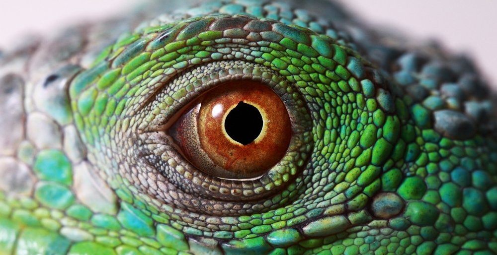
The lizard eye may be divided into the cornea near the front of the globe, and the orbital segment towards the back of the globe, with ‘globe’ referring to the eyeball itself. The cornea is made of tough, transparent tissue and acts like a window that controls and focuses the entry of light into the eye. The curvature of the cornea can be enhanced in lizards, alongside changes to lens spherity, through contractions of the anterior ciliary muscle. The retina is avascular and captures the light entering the eye and helps translate it into an image. It receives nutrition from the choriocapillaris. The conus papillaris is a feature of the reptile eye that projects into the vitreous cavity (the space between the retina and lens) to provide nutrition for the retina.
As with avian species, in the white part of their eyes lizards possess tiny bones called ‘scleral ossicles.’ They have 10-14 overlapping scleral ossicles that extend anteriorly. These give shape and stability to the globe and serve as support for the ciliary muscle. The ciliary body produces the fluid in the eye. Reptiles have a ciliary roll rather than the ciliary processes recognised in mammals and birds.
Lizards that possess eyelids, have a mobile lower lid, a relatively immobile upper lid, and a nictitating membrane or “third eyelid” which cleans the eye when they blink. The eye opens primarily through the downward movement of the lower eyelid, which contains a large cartilaginous plate. The exception to this is certain geckos and Ablepharine skinks which instead have fused eyelids that form a spectacle. Chameleons are another exception in that their eyelids constrict around the cornea and so there is more limited movement. Chameleons also have independent movement of each globe to enable them to fixate on prey species.
There are many differences between the eyes of different reptile species due to the different environmental niches that they inhabit. These differences in ocular anatomy and physiology can also be seen between different lizards. For example, nocturnal lizards possess large corneas and an almost spherical lens, and their pupil when dilated is hexagonal in shape to allow for a much larger pupil diameter. The depth of the globe is reduced compared to diurnal lizards to allow for greater irradiance on the retina. Whilst diurnal lizard retinas are only populated by cones, nocturnal lizard retinas possess cones that have developed morphological features of rods. The ellipsoid region of the cone photoreceptors of diurnal species contains oil droplets, which are believed to function as light filters and to contribute to colour differentiation.
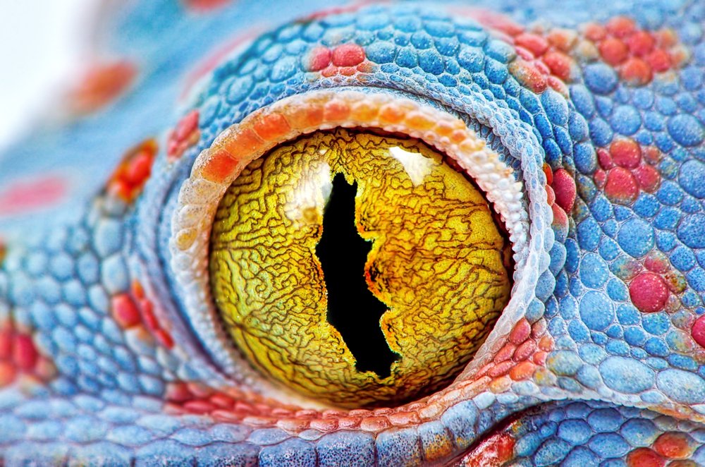
Another interesting difference is the polchorate (many pupiled) iris of some reptiles, such as the Tokay gecko (Gekko gecko). This multiple-pupiled feature is thought to affect refraction of light to help with understanding how far away a prey item is, prior to hunting it down.
There are two main ocular glands in lizards. These secrete fluid to lubricate and moisten the ocular surface. All lizards possess a Harderian gland which curls around the globe at its posterior aspect. Excluding chameleons and geckos, lizards also have a dorsotemporal lacrimal gland.
Many lizards also possess a ‘parietal eye’ which is quite forwardly positioned and often covered by a translucent skin scale. This structure is attached to an area of the brain called the pituitary gland. It plays a significant role in seasonal and diurnal physiological and behavioural cycles, such as reproduction, through light detection.
Common Disorders of the Lizard Eye
Conjunctivitis
Conjunctivitis is an inflammation of the eyelids, sometimes with concurrent eyelid swelling, and there can be mucoid ocular discharge, or the eyelids can become sealed together with dried discharge. This is commonly associated with bacterial infection in lizard species, and the author notes leopard geckos as being particular “offenders” in clinical practice.
Even if the eyelids are not sealed shut with dry discharge, conjunctivitis can impair the vision needed to see food items, and the infection and discomfort can also contribute to anorexia. Conjunctivitis can also be an important sign of septicaemia, not just localised infection of the eye alone.
Warm water compresses and gentle soaking can help as a first step in opening any sealed apertures. However, the eyelids should not be forced open to reduce the risk of trauma to the eye. A vet visit is recommended to assess the lizard for a correct diagnosis and thorough ophthalmological examination, and prescription of topical and/or systemic antibiotics will often be indicated. Flushing of the eye may also be warranted with warm saline and a sterile catheter by a trained professional, particularly in cases where retained shed or exfoliated skin are present.
Periocular Masses
Firm swellings around the eye that gradually grow and expand are the most common presentation of periocular masses reported in lizards. There can also be secondary trauma as the masses grow larger in size, and displacement and inflammation of the surrounding tissues as well as the growing mass itself can affect vision.
Bacterial lesions, fungal infections, tumours, infiltration of inflammatory cells leading to granuloma, and parasitic infection are all differentials.
Reptile pus is very thick and hard, and so it can be difficult to differentiate abscess from neoplastic mass, particularly just by feel or appearance. Some masses may also be both neoplastic and infectious/inflammatory. As such, the appropriate management for these must be based on an appropriate diagnosis. However, in most of these cases surgery is indicated to explore and resect/remove the mass that has developed, regardless of what it is.

Corneal trauma
As mentioned previously, the cornea is the clear outer part of the front of the eye. Damage or inflammation of the cornea can give it a cloudy or more opaque appearance. However, corneal trauma will often present as ocular discharge, excessive blinking of the affected eye(s) and holding it more closed than usual, and conjunctivitis.
Anything that rubs the surface of the eye could be a source of corneal trauma. For example, bedding caught in the eye, foreign bodies like grass seeds, or corners of sharp objects. Retained shed building up beneath the eyelids and being in contact with the surface of the eye is another possibility. For animals housed communally, trauma from conspecifics either accidentally or through fighting is also a risk.
Severity of trauma ranges from very minor and superficial to development of ulceration to perforation of the eyeball itself. Management is dependent on severity and underlying cause but may involve pain relief and topical medications, culture and sensitivity testing and appropriate antibiotics, tear replacement drops, or even surgery to repair the cornea. In all cases, any underlying husbandry issues should be identified and corrected.
Eye ulcers should be identified and appropriately treated promptly. They are known to be a painful condition in other species, and very severe ulcers can become deeper and lead to rupture of the globe. Uncomplicated corneal ulcers should heal within 1 week, though older animals may take slightly longer to heal.
Perforation seems less likely to occur in lizards with a spectacle as this acts protectively over the surface of the cornea. If there is damage to the structures within the eye, or intraocular infection occurs, then the prognosis of the eye is guarded, and it may need to be removed.
Keratitis
Though trauma is more commonly seen in lizard patients, non-traumatic corneal diseases can also be presented. Keratitis describes an inflammation of the cornea. Whilst this may be due to ulceration, it can also be noted in cases of infection and/or excessive provision of ultraviolet light.
Hypovitaminosis A
Although it is more commonly reported in turtles and tortoises, vitamin A deficiency is thought to be one of the most common causes of eyelid pathology in reptiles. This usually presents as swelling of the eyelids or periocular region, but there can also be ocular discharge and redness of the eye. There can be secondary bacterial infection associated with this condition, and there may be other disease processes going on internally due to the deficiency.
It is important to be aware of species variations when checking for eye redness as some reptiles, such as some varanid monitor lizards, have a red episclera (the “white” of the eye) which would be an indicator of ocular inflammation in a different species.
This condition is more commonly seen in younger animals and is typically associated with feeding a diet deficient in vitamin A.
Prevention of Ocular Conditions
Environmental temperatures and being within the Preferred Optimal Temperature Zone for a species is crucial for governing the rate of reptile physiological processes. Inappropriate thermal gradients and humidity will reduce any reptiles’ ability to maintain a healthy immune system to combat infection. A minimum of both the hot and cool end of any vivaria should be monitored continuously, ideally with a minimum-maximum thermometer to identify any fluctuations in temperature, particularly overnight. For vertical rise set-ups, the temperature gradient should expand in both the vertical and horizontal planes. Similarly relative humidity should be monitored constantly with a hygrometer, and any fluctuations around heat or lighting set-up should be noted and addressed, as necessary. It is important for the probes of these to be measuring at the level the inhabitant is within the set-up itself.
Low environmental relative humidity can lead to dehydration of the lizard and its skin. This, alongside affecting mechanical functions such as the properties of a gecko adhesive foot hairs, can be a contributing factor in difficulties with retained shed. In the author’s experience, retained shed around the head and eyes can be a particular issue in leopard geckos and result in secondary infections of the eye and, in more severe cases, periocular abscesses that require surgical removal. Conversely, inappropriately high humidity can have impacts on bacterial and fungal loads due to dampness of substrate or may make some substrates more adhesive. Ventilation should never be compromised to maintain correct humidity as this can also contribute to a build-up of airborne bacteria and fungal spores.
Within the set-up, care should be taken with substrate and paraphernalia choices so that anything included is appropriate for the species but with a minimal risk of adhesion to ocular surfaces or resulting in a traumatic injury. There should also be multiple refuge areas throughout the different humidity and heat gradients to allow inhabitants to remove themselves from ultraviolet light exposure if they deem it necessary. Appropriate environments for the species to facilitate shedding should also be provided. For example, a hide with damp moss that is misted daily should be provided for leopard geckos to help with removal of old sloughed skin. This also provides a necessary humid microclimate that allows the inhabitant to better regulate its humidity.
Providing a nutritionally balanced and appropriately supplemented diet is also crucial to ensure optimal body condition of the reptile and to reduce the risk of contributing vitamin deficiencies. All lizards should be fed appropriately for their individual species and age.
Conclusion
Ocular conditions are a frequent reason for lizards to require veterinary assistance. The conditions themselves can cause discomfort, and vision loss. Ocular conditions not only impact an animals’ ability to localise food but can also contribute to heightened levels of stress and consequent immunosuppression. Many ocular conditions in lizards can be linked to issues with housing and diet, and prompt identification and correction is needed. Eye trauma is a reason to seek immediate veterinary assistance.