By Dr Michaela Betts
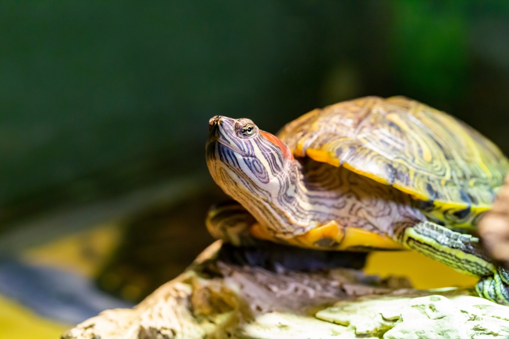
Aquatic turtles are relatively popular as pets in the UK. For many years, the Trachemys genus was most popular but today there is a wide variety of captive species available from the tiny Reeve’s turtle (Mauremys reevessii) to the larger common snapping turtle (Chelydra serpentina). Within the UK it is illegal to release non-native species and, since the introduction of the Invasive Alien Species legislation, it is an offence to breed, trade, or move Trachemys scripta subspecies, with keepers only being allowed to maintain owned animals for the rest of their natural lifespan. To keep these animals for their natural lifespan, it is important to be aware of not only their proper husbandry but to be familiar with common clinical signs and differential causes for disease processes.
Common Clinical Signs
Anorexia
Defined as a lack of appetite or intake of food, anorexia is not a disease but rather a non-specific result of an underlying pathological or physiological condition. Whilst it may be considered normal during certain times of the year, it is important to differentiate between physiological anorexia and reduced food intake due to an underlying disease process or inappropriate husbandry.
Inappropriate size and type of food offered, offering food at inappropriate times, suboptimal temperatures and humidity, inadequate exposure to ultraviolet lighting, poor water quality, lack of hiding areas, high stocking density, and a sudden change in diet are all potential husbandry issues that may contribute to anorexia. Any disease process or cause of stress to the animal could potentially present as anorexia so further investigations should be tailored towards identifying the cause and any negative consequences it has caused to the turtle as chronic anorexia can lead to fatty liver disease and nutritional and metabolic imbalances. Prompt investigation, diagnosis, and treatment of true anorexia and initiation of appropriate nutritional support can help to prevent the development of these negative consequences.
Shell Issues
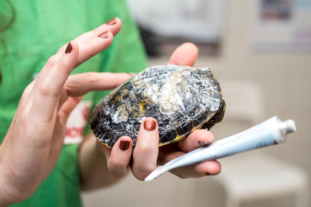
Algae growth may occur as green algae colonises the shell of aquatic chelonia. If the husbandry conditions are appropriate, this is generally of little clinical concern and can be periodically scrubbed off with a soft-bristle brush to keep the levels under control. However, excessive algal growth may reflect poor water quality and, if the shell is compromised, penetrate the shell surface. Water quality should be tested and husbandry practices reviewed to ensure appropriate.
White regions on the shell may be indicative of bacterial and/or fungal shell infection, healed shell lesions, mineral deposits, or exposed healed bone that is missing the overlying scutes. The “dry” form of septicaemia cutaneous ulcerative disease (SCUD) will often present as dry white patches on the carapace and plastron alongside signs of anorexia and lethargy. It has been suggested this Is partly due to a combination of desiccation of the scutes from basking and rehydration from immersion causing contraction and expansion of superficial unshed scutes, meaning that turtles continuously immersed or basking may be more prone to this issue. It is worth noting that parts of shed skin often remain attached to the limbs or body of aquatic turtles and may have a white appearance to them, and should not be confused with SCUD. Turtles should be promptly presented for veterinary assessment and treatment if infection is suspected as deeper infection of the bone and organs can occur if allowed to develop.
SCUD can occur in freshwater turtles of any age. Contributing factors are poor water quality, overcrowding, lack of ultraviolet lighting, temperature extremes, trauma and abrasive enclosure paraphernalia, lack of a dry basking area, and immunosuppression. These factors ultimately lead to an overwhelming bacterial infection which can lead to sepsis, organ failure, and death. The disease is associated with a variety of opportunistic bacteria that are part of the normal environmental biome and reptilian gastrointestinal tract and most commonly cause infection secondary to trauma that has broken the integrity of the skin or shell. The disease can cause pitting of the shell and forms circular lesions between the dermis and the scutes, causing them to ulcerate and in some cases to slough off, and may cause the loosening and sloughing of claws. The lesions typically start at the seams of the scute and spread inward, and may have a roughened or flaky appearance in the initial stages. Diagnosis is typically made based on the history and clinical signs. The prognosis depends on the severity and chronicity of the condition and treatment may take several months before healing is observed, with scar formation associated if bone infection is present.
Swelling
Swelling of the skin or the space beneath it can be a non-specific sign seen in Chelonia.
Generalised swelling beneath the skin may be seen if there is fluid build-up of the individual. This is generally associated with more serious underlying issues such as kidney, liver, or heart disease, low protein levels in the blood, or obstruction of the blood or lymphatic vessels. Bloodwork is usually indicated to rule out some of these underlying conditions. This should not be confused with obesity, where overweight individuals may deposit excessive fat in the axial and pre-femoral areas.
Swellings associated with joints may be seen with cases of articular or periarticular gout (a build-up of uric acid crystals in or around the joints), infection of the bone, joint, or soft tissue, parasites, metabolic bone disease, penetrating foreign body, or with trauma, including fractures and subluxations. Diagnostic testing and imaging are necessary for a definitive diagnosis of the underlying cause, and some of these causes require long-term management and support or may carry a poorer to guarded prognosis if they have progressed.
Ear Swelling
Swelling over one or both ears is a presenting complaint in chelonians. Unlike mammals, the turtle ear is a simple structure that lacks an open external ear canal and is instead covered by a large scale known as the ‘tympanic scute’. The middle ear is a large tympanic cavity bordered by the eardrum, which is in direct contact with this overlying skin. The most common underlying cause of ear swelling in captive aquatic and semi-aquatic turtles seems to be abscessation. These can cause severe internal and external deformities if left to grow. Whilst the abscesses can be sterile, the majority are associated with bacteria and form a thick, caseous plug of purulent material within the middle ear. However, other possible causes for ear swelling are parasites underneath the skin, sebaceous cysts, haematomas (a solid swelling of clotted blood), and mycobacterial skin infections.
The cause of ear abscessation is not completely understood, but studies have suggested vitamin A deficiency may predispose turtles to develop the condition. It is suggested this is due to the impact on the immune system and the key role vitamin A plays in the normal maintenance of healthy skin. Secondary infection is typically associated with ingestion of contaminated water or an overgrowth of opportunistic bacterial in the mouth that then ascend the Eustachian tube that connects the middle ear to the throat. Appropriate temperature, water quality, and dietary supplementation are crucial in reducing the risk of developing this condition.
Successful treatment of ear abscesses requires surgical removal of the purulent material and appropriate antimicrobial therapy. Most turtles with ear abscesses surgically removed respond well to treatment and heal completely. However, the issue can recur if underlying vitamin deficiencies are not addressed, if there is an additional infection of the bone present, and if the infective material is not completely removed at the time of surgery.

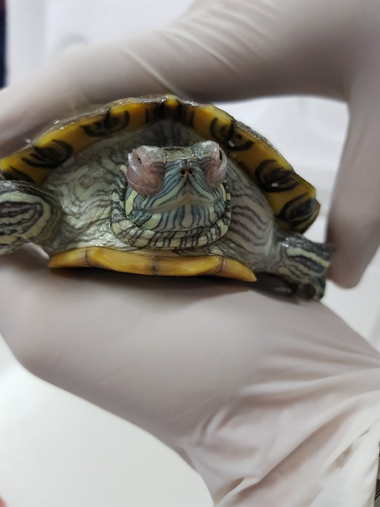
Swollen Eyes & Ocular Discharge
Ocular conditions are often indicators of a systemic infectious processes rather than a primary eye issue. Discharge from the eyes and swelling around the eyelids can be seen in association with upper respiratory tract disease, ocular irritants or trauma, viral or bacterial diseases, and low levels of vitamin A. Sea turtles lack lacrimal ducts and so produce a clear ocular discharge normally.
Hypovitaminosis A is a very common underlying aetiology of swollen eyes in turtles, and it results in a build-up of epithelial cells associated with the eyelids. Unfortunately, it is usually a result of long-term inappropriate diet and supplementation. The swelling can range from mild swelling of the eyelids to severe swelling with conjunctivitis to the extent that the individual is blinded. Secondary infection is commonly seen with this condition that requires antibiotic therapy. Successful treatment involves correcting the diet and multivitamin/multimineral supplementation, and ensuring that the rest of the environment is appropriate in terms of temperatures, basking area, lighting provision, and water quality. In some cases, particularly those where the turtle is non-visual due to the swelling, hospitalisation may be necessary to commence appropriate supportive care and medication and to treat the eyes as necessary.
Herpesvirus can affect all ages and species of Chelonia, and there are at least four different strains reported. In aquatic turtles, it has been reported in various emydid species. Whilst it can present as ocular discharge signs, in aquatic species, it more often presents as weakness, nasal discharge, lethargy, stomatitis, hepatitis (liver inflammation), and sudden death. Viral detection is performed with polymerase chain reaction (PCR) detection of viral DNA. Appropriate quarantine measures should be in place to help avoid introducing the virus to a collection, and the mixing of different species of turtle should be avoided.
Erythema
Pink or red discolouration of the skin or beneath the shell is often an indicator of an infectious process such as a bacterial skin infection or, in some cases, serious septicaemia (blood poisoning by bacteria), or sometimes with hypovitaminosis A. Though it may seem relatively benign, the underlying cause can be a very serious clinical problem and, in most cases, warrants further investigation to identify and treat the underlying problem. Keepers should be familiar with the colouration of their turtles to identify if erythema is developing. Many juvenile freshwater species naturally have a reddish colouration to their plastron that fades as they age, and this should not be mistaken for erythema.
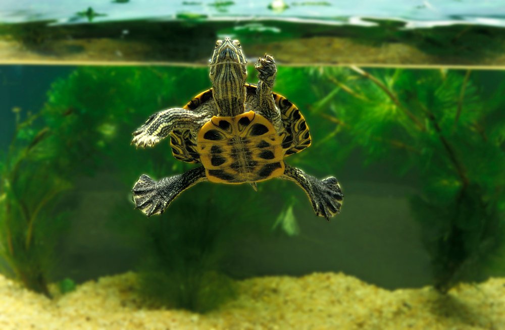
Abnormal Submersion
Some health issues may present as abnormal floating at the water surface (front-to-back or uneven from side-to-side) or an inability to submerge. Whilst the latter is more commonly displayed in sea turtles, these symptoms can be a sign of pneumonia as the pneumonic lung becomes heavier, gas accumulation within the gastrointestinal tract or coelomic cavity, or from any sort of unilateral mass within the individual such as a tumour, abscess, or foreign body. Diagnostic imaging should be pursued with a reptile-savvy vet to identify potential underlying factors, and next steps based on the findings.
In Conclusion
Whilst they can be rewarding to keep, aquatic turtles require a lot of work to be properly maintained. An understanding of appropriate temperature, lighting, water quality, basking area, and dietary supplementation is crucial in reducing the risk of developing health issues, as well as the many species-specific differences between them. As with other reptile species, signs of illness can be vague or non-specific compared to more commonly pet mammals, and keepers should be aware of clinical signs to look out for and the potential underlying causes.