An introduction to husbandry and commonly occurring health problems
By Dr Michaela Betts
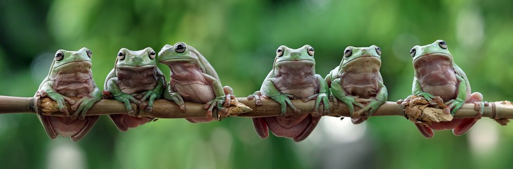
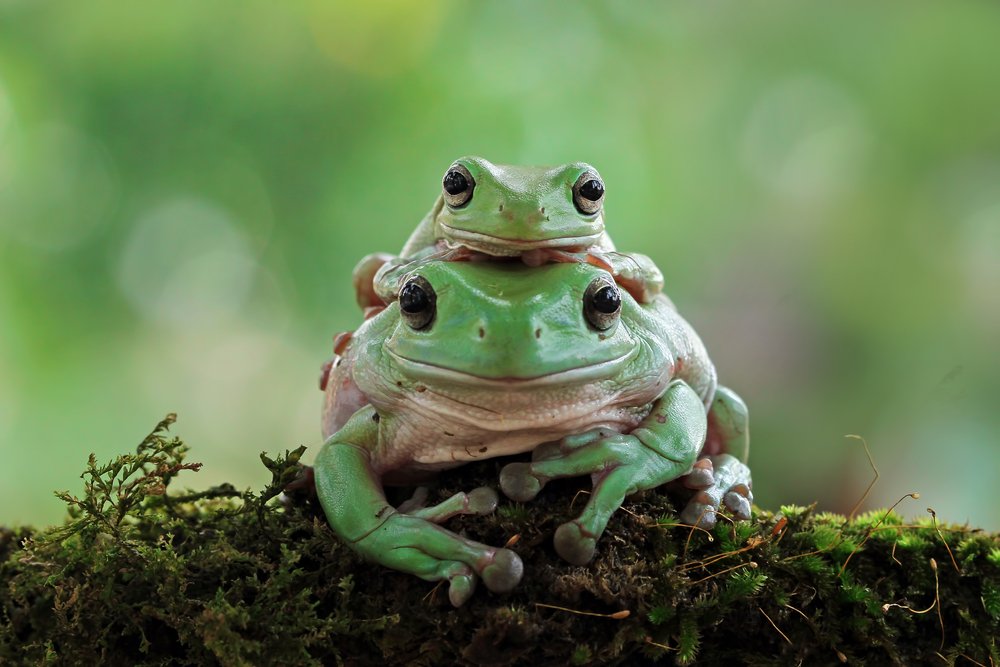
Captive frog and toad species are increasingly popular in the pet trade, while also being important from an ecological perspective. Of the estimated 100,000 pet frogs and toads in the UK in 2018, the White’s tree frog (Litoria caerulea) was one of the most-commonly kept species. Dr Michaela Betts, an exotic animal veterinarian working in Suffolk, has seen this reflected in clinical practice with the White’s tree frog being the most common frog species presented.
Litoria caerulea
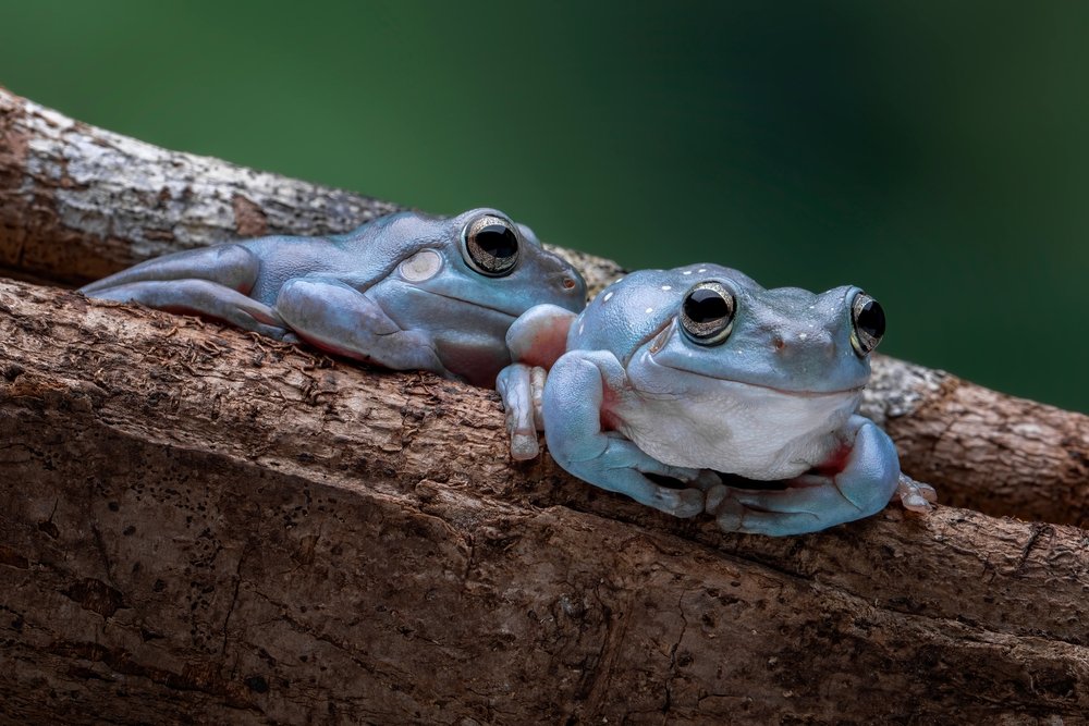
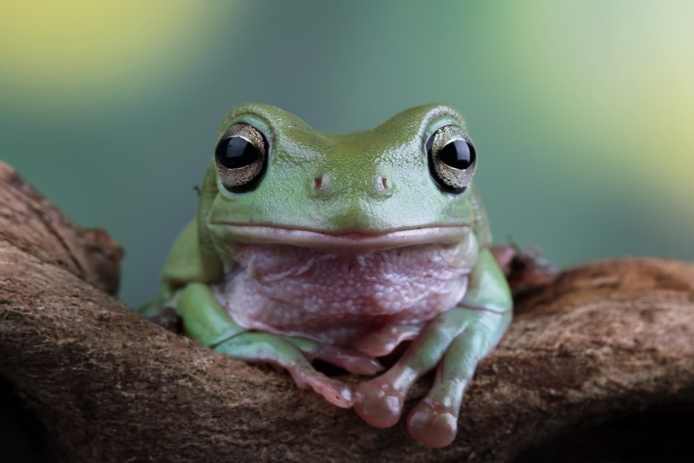
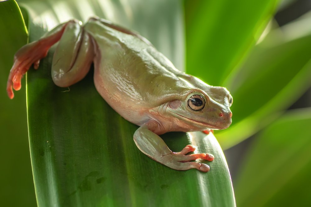
The White’s tree frog, also known as the “dumpy tree frog” is an Australasian tree frog indigenous to Indonesia and Australia. They are a nocturnal, arboreal species with a wide variation in colouration, from rusty brown, to green, to blue-tinged. They are medium-large with adults typically measuring between 50-110mm in length.
Housing
In the wild, White’s tree frogs thrive in a variety of habitats but are more commonly found in humid coastal regions and require an environment with appropriate heat, humidity, and ultraviolet (UV) lighting. They do well in set-ups constructed from non-abrasive materials that do not leach chemicals, like glass vivariums, with one to three frogs being able to thrive in a 45x45x60 enclosure.
This species should have a daytime temperature gradient of 24-29°C with a 35°C basking spot, reducing to 18-21°C overnight. This can be achieved with a thermostatically controlled heat mat on the side of the vivarium, a small basking bulb, ceramic heat emitter or a combination of these. However, any ceramic heat emitter needs to be kept far enough away from the highest basking spot to prevent thermal burns or inadequate humidity. Temperatures should be measured at both the hot and cool ends of the set-up using a minimum/maximum thermometer to ensure there are no unknown fluctuations throughout the day and night. It is recommended to have a slightly lower humidity and daylight temperature during the winter months.
Humidity should be maintained between 60-70%, with daily misting and constant access to a sizeable but shallow water dish. A hygrometer is essential to ensure relative humidity is always maintained. Water should be changed daily. It is recommended to either use spring water, or chlorinated water that has been aerated in an open container for 24-48 hours to allow the chlorine to dissipate.
Ultraviolet lighting is important to provide a regular photoperiod and enable efficient vitamin D3 production. A low percentage UV light for a 10-12 hour photoperiod is advised. Keepers with a means of measuring UV index should be aiming for a range of 0.7-1.6 for this species. Additionally, UV bulbs need to be replaced roughly every 6-12 months as the UV-B emitted reduces over time.
Opaque refugia must be included in the enclosure to provide hiding spots and visual security. White’s tree frogs benefit from a perching spot at least the diameter of their body such as a clay pot or tree limb.
Housing challenges
Maintaining a high enough humidity alongside good provision of UV-B can be difficult. Equally, ensuring that substrate remains damp but does not become water-logged can be a challenge.
If the substrate becomes sodden, a build-up of ammonia often develops which can lead to health issues that require veterinary attention. Keepers can use a false bottom made of expanded clay balls or koi fitter crate with a layer of fly screen to separate the water layer and the substrate. A mixed substrate that utilises soil, bark chips, coir and moss is often preferred, with a final layer of leaf litter on top such as oak or beech.
Diet
White’s tree frogs are insectivores and, for optimal nutrition, should be fed a variety of appropriately sized gut-loaded invertebrates – primarily insects. Commonly fed insects are locusts, crickets, dubia roaches and the occasional waxworm treat. As an arboreal species, insects that attach to vegetation or furnishings at the level of the frog are preferable. Food items should be dusted with a calcium and multivitamin supplement, usually once weekly for adults and twice weekly for juveniles. Similarly, juveniles should be fed daily while adults can be fed two to three times a week. It is best to feed during peak activity levels, in the evening, to ensure that food is eaten whilst the supplement coating is still on.
Nutritional disease
Obesity is common with this species. It is often associated with a cloudy appearance to the eyes as the excessive dietary fat results in a lipid keratopathy, along with an enlarged appearance to the coelom due to fat deposition. In White’s tree frogs specifically, obese individuals may also deposit fat in the crests over their eyes which can further impede vision if they become large enough. As with other species, increasing activity levels and reducing their food is the recommended course of action. Unfortunately, there is currently no specific treatment for the ocular changes, so it’s advised to limit calorific intake to prevent progression of the lesions. Analgesia may be added if the condition seems to be causing discomfort.
Metabolic bone disease is another common condition, often due to nutritional secondary hyperparathyroidism. This typically affects juvenile frogs and is associated with inadequate UV-B provision, inadequate oral calcium supplementation, low dietary calcium/phosphorus ratio, inappropriate heating or a combination of these factors. Inappropriately high phosphorus or fluoride levels in the water has also been associated with the condition. Review of the diet, water source, UV-B lighting and heating should therefore be carried out if a diagnosis is suspected or confirmed by x-ray imaging. Severe cases often require analgesia, as well as additional calcium supplementation and repeat x-rays at regular intervals to assess for improving bone density. Obvious clinical signs include skeletal deformities or fractures. Although less-specific symptoms are general weakness, accumulation of gas in the gastrointestinal tract and a bloated appearance, cloacal prolapse, and death.
Handling
Clean, powder-free gloves, moistened with dechlorinated water should be used for handling any anuran, but handling should be kept to a minimum. Direct contact with human skin should be avoided due to the semipermeable nature of amphibian skin. Because of the comparatively more tolerant nature of this species, White’s tree frogs are likely to be handled more often in the home environment than other anurans. Unfortunately, this brings with it the risk of contaminants such as chemicals or bacteria plus stress – which can cause immunosuppression and may lead to the development of secondary disease processes.
Quarantine
Appropriate quarantine measures for any newly acquired amphibian are vital for preventing the introduction of disease into a collection. Whilst captive-bred in large numbers, wild-caught White’s tree frogs can still be found in the pet trade. The amphibian pet trade itself has been implicated in the spread of infectious diseases, most notably Batrachochytrium dendrobatidis and ranaviruses. Therefore, adequate research of the source of your frog and the implementation of appropriate quarantine procedures are of particular importance.
One size does not fit all. The length of a quarantine period and choices around infectious disease screening of your new tree frog will vary depending on the source and history of the individual or group. Ideally, a quarantine set-up should be kept in a dedicated area away from other amphibians with footwear and protective clothing for that location only. Dedicated tools and equipment, handwashing and new gloves should be kept for quarantine populations. Plus tools and equipment used between different quarantine enclosures should be thoroughly disinfected. Paper towel substrate can suffice for this species and allows observation of faeces and ease of cleaning in quarantine. However, care must be taken to maintain adequate humidity.
High-risk animals are those from an unknown source or with poor biosecurity measures, as well as animals with an unknown health history or that appear unwell. Sharing a single room or water system with unwell amphibians or being with species from different geographical locations or sources can also put animals at high risk. Conversely, lower risk animals are those from a source with good biosecurity practices, who have a complete health history and have been kept in a single species facility or are from a population at a mixed species facility which had been isolated long-term before acquisition.
Regardless of risk level, it is recommended to test any new frog for Batrachochytrium due to the significance of the disease and its implications for both the collection and the individual frog. While it is recommended to test for this at least once in low-risk animals, higher risk animals should ideally be tested two or three times over their quarantine period. Faecal examination at the start and end of a quarantine period is also recommended to ensure appropriate identification and treatment of any parasites.
The recommended quarantine length varies depending on the risk. A minimum of 30 days should be adhered to, though 60-90 days is more appropriate for high-risk animals, and 90 days should be the minimum if wild-caught. However, if there are unexplained deaths, a significant pathogen is identified, new animals enter their quarantine area, or there are health problems or treatments in a quarantined population, then the period should be extended.
Fungal disease
The most significant fungal pathogen of anurans is the causative agent of chytridiomycosis, Batrachochytrium dendrobatidis. Chytrid fungi are ubiquitous in moist and aquatic environments and chytridiomycosis is transmitted by direct contact with infected individuals, contaminated water or through human or mechanical vectors. Larval anurans are typically subclinically infected, with the infection occurring in the mouthparts. However, in postmetamorphic anurans, infection occurs in the keratinising stratified squamous epithelium of the skin and has a very high mortality rate. Clinical signs include lethargy, oedematous swelling, abnormal posture and loss of the ability to right itself, abnormal behaviour such as a loss of flight or fight response when handled, dehydration, dysecdysis and hyperaemia of the skin. The infection disrupts the affected skin’s ability to transport electrolytes across it, so sadly these individuals pass away from electrolyte depletion. This change to the skin can also lead to secondary bacterial infections and so should always be considered as a potential underlying cause of ‘red-leg’.
Diagnosis can either be made by histopathology at post-mortem or by polymerase chain reaction (PCR) testing from skin swab samples of the inner thighs, underside of hindlimb digits and webbing, plus drink patch in live frogs.
Various treatments have been reported for chytridiomycosis and there is no single protocol suitable for all species and life stages. Medical bathing for 11 consecutive days seems to be the favoured treatment method for this species at present.
As the organism can persist for a long time in moist environments, affected animals should be kept in a clean, sparse, disinfected and easily cleaned enclosure. The permanent enclosure should be deep cleaned and disinfected prior to the return of successfully treated individuals. Strict biosecurity measures also need to be followed during this period to reduce the risk of spreading the infection. The effectiveness of any treatment should be monitored with post-treatment repeat PCR testing, with the current recommendation being three PCR tests over a 14-day post-treatment period.
Bacterial disease
Many bacteria make up the normal flora of the skin but can become pathogenic if there is trauma or immunosuppression from inappropriate husbandry, viral infection or fungal infection. Common potentially pathogenic bacteria include Aeromonas, Pseudomonas, Escherichia coli, Mycobacterium spp., and Chlamydophila.
The classic appearance of a frog suffering from a bacterial dermatosepticaemia is ventral erythema and petechial haemorrhage which gives the colloquial ‘red-leg’ syndrome its name, often alongside ulceration, erosion, sloughing and necrosis of the skin. However, there are many other clinical signs associated with bacterial infections that are more non-specific, like anorexia, lethargy, abnormal gait, and coelomic effusion. White’s tree frogs can also develop a cloudy appearance to their eyes due to corneal oedema secondary to bacterial infection.
Investigations should include skin swabs for culture and sensitivity and impression smear, skin scrapes for cytology and wet mount and possibly swab samples for PCR testing if Mycobacterium is suspected. Mycobacterium is of significance due to its zoonotic potential. Treatment involves parenteral antibiotics, ideally based on culture and sensitivity testing. Depending on underlying causes, treatment of primary infections, correction of water quality or husbandry may also be indicated.
Parasitic disease
A wide variety of parasites have been documented in captive anurans and can be investigated through faecal sampling. Fresh faeces can usually be collected from moist paper towel in a clean container and by target-feeding the individual.
Many metazoan and ciliate protozoa are commensals in anurans, and bacteriology is often of little clinical significance even if there is a pure growth of a single isolate. Therefore, the decision to enact treatment is dependent on the type of organism identified, the parasite load, and on associated clinical signs such as ill thrift, anorexia, diarrhoea or prolapse. In all positive cases, isolation of the affected individual and measures to reduce the risk of re-infection from contaminated material should be pursued.
Potentially pathogenic protozoa include Entamoeba ranarum, Microsporidium spp., Chloromyxum spp., Myxidium immersum, Eimeria, Isospora, and Myxobolus hylae. Whilst some of these primarily affect the gastrointestinal tract, others can have impacts on other organs or the striated muscles.
Nematodes, cestodes and trematodes can also be found in anurans, with the latter typically existing in larval stages. With these, the clinical signs are usually minimal. However, larvae can sometimes encyst within the dermis, causing large focal pathology that will require surgical intervention by a veterinarian to remove the worm(s). A notable nematode of this species is the lungworm Rhabdias, which can cause respiratory symptoms. The larvae or embryonated ova of this parasite can be found on faecal examination and sometimes by microscopy of an oral smear.
What next?
Research into appropriate husbandry and the identification and treatment of disease processes in amphibians is still a growing area. Even among exotic veterinarians, amphibians will typically only make up a small percentage of animals presented. Although there is still much to learn, there is a growing understanding of how to appropriately house and treat these species at home and in clinical practice. Appropriate quarantine and husbandry with prompt identification, isolation, and treatment of diseased individuals are key factors in maintaining a healthy collection.